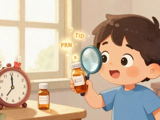


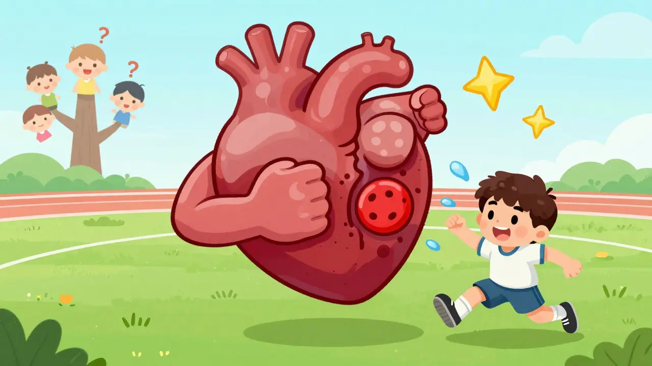
When your heart muscle weakens or stiffens, it doesn’t just feel like fatigue-it can be life-threatening. Cardiomyopathy isn’t one disease. It’s a group of conditions that attack the heart muscle directly, making it harder for your heart to pump blood. The three main types-dilated, hypertrophic, and restrictive-each behave differently, need different tests, and require totally different treatments. Most people have never heard of them until they or someone they love gets diagnosed. And that’s the problem: these conditions are often missed, misdiagnosed, or delayed because they don’t look like a typical heart attack.
Dilated Cardiomyopathy: The Heart That Swells and Fails
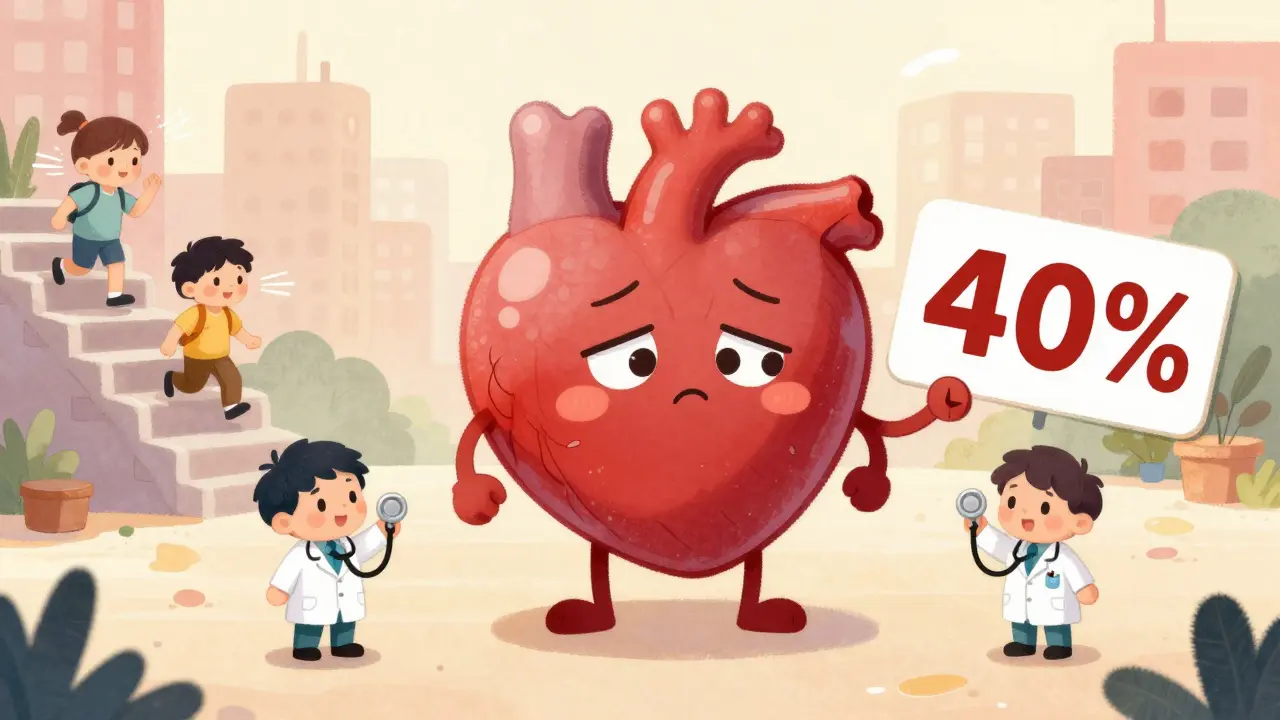
Dilated cardiomyopathy (DCM) is the most common type, making up over half of all cases. Imagine your heart’s main pumping chamber-the left ventricle-stretching out like an overinflated balloon. The walls get thin, the chamber grows too large, and the muscle loses its strength. By the time it’s diagnosed, the ejection fraction (how much blood it pumps out with each beat) often drops below 40%. A healthy heart pushes out 55-70%. This one struggles to hit 30%.
Why does this happen? Sometimes, it’s genetic. Mutations in genes like TTN, LMNA, or MYH7 can be passed down, even if no one in the family ever had symptoms. About one in three people with DCM have a family history. Other causes include long-term heavy drinking (more than 80 grams of alcohol a day for five years or more), viral infections like coxsackievirus, or damage from chemotherapy drugs like doxorubicin. Autoimmune diseases like sarcoidosis can also trigger it.
People with DCM often feel winded climbing stairs, swollen in the legs, or like they can’t catch their breath lying down. The diagnosis starts with an echocardiogram: if the left ventricle is bigger than 55 mm in men or 50 mm in women, and the ejection fraction is under 40%, it’s DCM. Cardiac MRI is now standard to check for scar tissue or inflammation. Genetic testing is recommended if there’s a family history-especially since early detection can save lives in relatives.
Treatment isn’t about fixing the heart back to normal. It’s about slowing the decline. Guideline-directed medical therapy (GDMT) includes drugs like sacubitril/valsartan (a newer alternative to old ACE inhibitors), beta-blockers, and SGLT2 inhibitors originally made for diabetes. These reduce hospitalizations and cut death risk by 30% over three years. For some, an implantable defibrillator is needed to prevent sudden cardiac arrest. In advanced cases, a heart transplant may be the only option.
Hypertrophic Cardiomyopathy: The Heart That Grows Too Thick
If DCM is the heart stretching out, hypertrophic cardiomyopathy (HCM) is the heart growing inward-too thick, too stiff. The muscle, especially in the septum (the wall between the left and right ventricles), becomes abnormally thick-15 mm or more-without any high blood pressure or valve disease to explain it. This isn’t the kind of thickening you get from lifting weights. This is chaotic, disorganized growth at the cellular level.
HCM affects about 1 in 500 people, but many go undiagnosed. It’s the #1 cause of sudden cardiac death in young athletes under 35. In the U.S., it accounts for 36% of those tragic cases. The problem? The thickened muscle can block blood flow out of the heart-this is called obstructive HCM, and it happens in 70% of cases. When that happens, even mild exercise can cause dizziness, chest pain, or fainting.
Genetics play the biggest role. Around 60% of cases come from mutations in genes like MYH7 or MYBPC3, inherited in an autosomal dominant pattern. That means if one parent has it, each child has a 50% chance of inheriting the gene. A 17-gene panel test costs $1,200-$2,500 in the U.S., and it finds the cause in about 60% of cases. Even if the test is negative, the diagnosis still stands if the wall thickness is clear on echo or MRI.
Diagnosis is straightforward: echocardiogram shows the thickened wall, and stress tests or cardiac MRI confirm if there’s obstruction. A gradient of 30 mmHg or more at rest means it’s obstructive. Treatment starts with beta-blockers-70% of patients feel better. For those with severe obstruction, disopyramide (a heart rhythm drug) can help. But for the worst cases, there are two surgical options: septal myectomy (removing part of the thickened muscle) or alcohol septal ablation (injecting alcohol to shrink the area). Both improve symptoms in 85% of patients. In 2022, the FDA approved mavacamten (Camzyos), the first drug made specifically for obstructive HCM. It reduces the blockage by 80% and cuts symptoms dramatically.
What’s scary is how easily HCM can be missed. Athletes are routinely screened, but many aren’t. A young person who collapses during a game? Often, it’s HCM. And if you have a family member who died suddenly before age 50, you should get checked-even if you feel fine.
Restrictive Cardiomyopathy: The Heart That Won’t Fill
Restrictive cardiomyopathy (RCM) is the rarest-and most misunderstood-of the three. It doesn’t make the heart big or thick. Instead, it makes it stiff. The muscle is normal in size, even strong in squeezing power, but it can’t relax enough to fill with blood. Think of it like a balloon that’s been glued to the inside of a jar-it can’t expand properly.
RCM affects only 2.5-5% of cardiomyopathy patients. Its hallmark is a normal ejection fraction (over 50%) but terrible diastolic function. Echocardiograms show a fast, sharp “E wave” (early filling) with a very short deceleration time (under 150 ms). The atria get huge because blood backs up into them. Pulmonary pressures rise, causing fluid in the lungs and swelling.
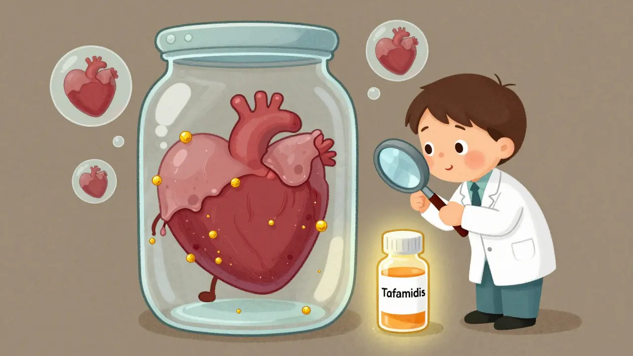
Unlike DCM or HCM, RCM isn’t usually genetic. It’s caused by other diseases that invade the heart. Amyloidosis is the biggest culprit-60% of cases. That’s when abnormal proteins build up like sludge in the muscle. Sarcoidosis, hemochromatosis (too much iron), and rare storage diseases like Fabry disease make up the rest. Cardiac MRI is key here: it shows a unique pattern of late gadolinium enhancement-not in the arteries, but scattered through the muscle. Extracellular volume over 35% means fibrosis is widespread.
Diagnosing RCM is tricky. The biggest pitfall? Mistaking it for constrictive pericarditis, where the sac around the heart gets stiff. The treatments are completely different. One needs surgery to remove the sac; the other needs to treat the underlying disease. That’s why biopsy is often needed-especially to confirm amyloidosis. A biopsy shows the waxy amyloid deposits under a microscope.
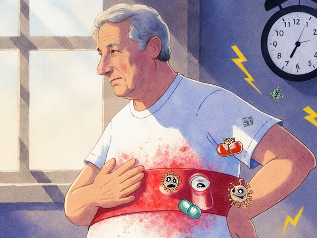
Treatment depends entirely on the cause. For light-chain amyloidosis, drugs like daratumumab (a cancer immunotherapy) are used. For hemochromatosis, regular blood removal (phlebotomy) helps. Tafamidis, a drug that stabilizes the amyloid protein, costs $225,000 a year in the U.S., but it improves walking distance by 25 meters in six months. Survival rates are poor-only 30-50% at five years, depending on the cause. That’s why early detection matters more than ever.
Why Classification Matters
Doctors used to group all heart muscle diseases together. Now, we know better. The American Heart Association and European Society of Cardiology updated their guidelines in 2020 and 2021 to separate primary cardiomyopathies from secondary causes like coronary artery disease. That’s critical. If your heart failure is caused by clogged arteries, you need stents or bypass-not the same drugs as someone with genetic DCM.
And here’s the kicker: up to 15-20% of DCM cases are misdiagnosed because no one checked for blockages first. That’s why a coronary angiogram is now standard before labeling someone with idiopathic DCM. Similarly, RCM gets mislabeled as heart failure from high blood pressure-when it’s actually amyloidosis.
The future is precision medicine. CRISPR gene editing is entering trials for HCM (VERVE-201 trial targeting MYBPC3 gene). Polygenic risk scores will soon predict who’s likely to develop HCM before symptoms appear. Genetic testing is becoming cheaper, faster, and more accurate. And new drugs like mavacamten and tafamidis are turning once-deadly conditions into manageable ones.
What You Should Do If You’re Concerned
If you have unexplained shortness of breath, fainting spells, chest pain during exercise, or a family history of sudden cardiac death before age 50, get evaluated. Start with an echocardiogram. If it’s abnormal, ask for a cardiac MRI and genetic counseling. Don’t wait until you’re in heart failure.
Even if you feel fine, if a close relative had HCM or died suddenly, get screened. Many people live with HCM for years without symptoms-until they don’t.
For RCM, if you have a known systemic disease like amyloidosis, sarcoidosis, or hemochromatosis, ask your doctor to check your heart. It’s not enough to treat the lungs or liver-you need to know if your heart is involved too.
Cardiomyopathy isn’t a death sentence anymore. But it won’t get better if you don’t know what you’re dealing with. The right diagnosis leads to the right treatment-and sometimes, it saves your life.
What’s the difference between dilated and hypertrophic cardiomyopathy?
Dilated cardiomyopathy (DCM) means the heart chamber is enlarged and the muscle is weak, leading to poor pumping. Hypertrophic cardiomyopathy (HCM) means the heart muscle is abnormally thick, especially in the septum, which can block blood flow. DCM reduces ejection fraction; HCM often keeps it normal but impairs filling. DCM is often caused by alcohol, viruses, or genetics; HCM is almost always genetic.
Can restrictive cardiomyopathy be cured?
There’s no cure for restrictive cardiomyopathy itself, but treating the underlying cause can improve outcomes. For example, amyloidosis can be treated with daratumumab or tafamidis; hemochromatosis with blood removal. If caught early, these treatments can stabilize the heart and extend life. In advanced cases, a heart transplant may be the only option.
Is hypertrophic cardiomyopathy hereditary?
Yes. About 60% of HCM cases are caused by inherited gene mutations, usually in sarcomere proteins like MYH7 or MYBPC3. It follows an autosomal dominant pattern, meaning each child of an affected parent has a 50% chance of inheriting the gene. Even if symptoms don’t appear right away, genetic testing can identify carriers and allow for early monitoring.
What tests are used to diagnose cardiomyopathy?
Echocardiogram is the first test for all types. Cardiac MRI helps detect scar tissue, amyloid deposits, or muscle thickness. Genetic testing is recommended for DCM and HCM with family history. For RCM, endomyocardial biopsy may be needed to confirm amyloidosis or sarcoidosis. Blood tests like NT-proBNP help assess heart strain.
Can you live a normal life with cardiomyopathy?
Many people do-with proper treatment. DCM patients on guideline therapy often live 10-15 years or more. HCM patients with non-obstructive disease have near-normal life expectancy. RCM prognosis depends on the cause; amyloidosis has a shorter outlook, but new drugs are improving survival. Lifestyle changes, regular follow-ups, and avoiding triggers like alcohol or intense exercise (in HCM) make a big difference.
10 Comments
Wow, this post is a game-changer. I’ve been working with cardiac patients in Mumbai for over a decade and I can’t tell you how many times I’ve seen DCM mistaken for asthma or anxiety-especially in young women. The part about genetic testing being recommended even if no one in the family showed symptoms? That’s huge. My sister-in-law was diagnosed with DCM after her cousin died suddenly at 28, and she had zero symptoms. Genetic screening saved her life-and now we’ve tested three other relatives. If you have unexplained fatigue or swelling, don’t wait. Get that echo. It’s not dramatic, it’s just smart.
Also, SGLT2 inhibitors for heart failure? I still get excited about that. Originally for diabetes, now it’s cutting hospitalizations by a third? That’s like finding a new vaccine for heart disease. And the fact that mavacamten just got FDA approval for HCM? Huge. We’re moving from just managing symptoms to actually targeting the root cause. This is precision medicine in real time, not sci-fi.
And RCM? People think if the ejection fraction is normal, everything’s fine. Nope. That stiff heart is silently drowning in fluid. Amyloidosis is terrifying because it looks like old age until it doesn’t. I had a patient who thought her joint pain was arthritis-turned out it was light-chain amyloidosis, and her heart was already failing. Biopsy saved her. Don’t let normal numbers fool you.
And yes, if your grandparent died suddenly before 50? Get screened. Even if you’re fit. Even if you’re young. I’ve seen athletes collapse because no one thought to check for HCM. It’s not about being paranoid-it’s about being prepared. Your heart doesn’t ask for permission before it fails.
While the clinical overview is commendably thorough, I must take issue with the casual tone adopted throughout. The use of metaphors such as ‘overinflated balloon’ and ‘glued to the inside of a jar’-while perhaps intended for accessibility-undermines the scientific rigor expected in medical discourse. Cardiomyopathies are not literary devices; they are complex, genetically mediated pathophysiologies requiring precise terminology. The reference to ‘cheap’ genetic testing is particularly concerning, as the cost-benefit analysis of population-level screening remains contentious in peer-reviewed literature. A more restrained, evidence-based presentation would better serve the intended audience of clinicians and informed patients alike.
So… you’re telling me the heart’s just a lazy balloon that got too big, a gym rat that got too bulky, or a balloon stuck in a jar? And we’re supposed to be impressed because we now have a $225k drug to un-stick it?
Look, I’m glad we’re finally catching up. But honestly? This feels like medicine playing whack-a-mole with biology. We fix one thing, another breaks. We give a drug that targets a gene mutation, then someone gets a rare side effect that turns their liver into a brick. We’re treating symptoms like they’re puzzles to solve, not systems to understand.
Meanwhile, people are still dying because their doctor didn’t think to ask about family history. Or they couldn’t afford the echo. Or the MRI waitlist is six months. We’ve got the science. We just don’t have the will.
Get an echo if you have unexplained shortness of breath or family history of sudden death. That’s it. No fluff. No drama. Just do it.
OMG this is like a Netflix documentary but real and I’m crying. I didn’t know my uncle’s ‘heart attack’ at 42 was actually HCM. He was a marathon runner. He looked healthy. He never complained. And now I’m scared to run up the stairs. But also-thank you. This is the kind of info we NEED. My cousin in Lagos just had a baby and I’m already texting her to get her baby’s heart checked. Genetics don’t care about borders. This is global.
Also, tafamidis costs more than my car. But if it gives me 6 more months with my mom? I’ll sell the car. I’ll sell my soul. This is life or death. And we’re talking about it like it’s a textbook. It’s not. It’s our families.
So we’ve got a heart that’s too big, too thick, or too stiff… and we’re calling it three different diseases. But what if it’s just one disease? What if the heart’s just… tired? Like, evolution didn’t design us to live 80 years with processed food, stress, and zero sleep? Maybe DCM, HCM, RCM aren’t separate-they’re just different ways the heart gives up.
We spend billions on gene editing and $200k drugs… but what if the real cure is less screen time, more sleep, and no more soda? I’m not saying we scrap the science. I’m saying we forgot to ask the most basic question: Why is this happening to so many people now?
And why is the answer always ‘more testing’ and never ‘less suffering’?
So like… if your heart is a balloon, who’s the one blowing it up?? 😳 Like… is Big Pharma doing this?? Like… did they make a virus?? I’m not saying it’s a hoax but… why is this only happening now?? And why is the cure always $$$?? 🤔 #CardiomyopathyConspiracy #WakeUpSheeple
This article is too long and too soft. In India we don’t have time for metaphors. If your heart is failing, get tested. If your uncle died young, get tested. If you can’t climb stairs, get tested. No one cares about your emotional journey. The heart doesn’t care if you’re sad. It just stops. So stop wasting time and go get an echo. Now.
You people talk like your heart problems are special because you have fancy machines. In Nigeria we don’t have MRI or genetic tests. We have prayers and traditional healers. But guess what? We still know when someone’s heart is failing. We see it in their face. We feel it in the silence after they stop laughing. You think your $225k drug is saving lives? We save lives with family, food, and faith. Stop acting like your science is the only truth.
ok so i just read this whole thing and i’m like… wait… so if i have a family member who died suddenly… and i feel kinda tired… and i’m 32… should i just… go get an echo?? like… right now?? because i think i might have… like… a thing?? and also i think i read somewhere that stress causes this?? but i don’t know if that’s true?? and also i think my doctor might be lying to me?? i mean… they’re just trying to sell me meds right??