


When a doctor orders a brain MRI, it’s usually because something doesn’t add up - a sudden headache, memory lapses, numbness, or vision changes. But what you see on the scan isn’t always what it seems. A bright spot on an MRI doesn’t automatically mean a tumor. A dark area isn’t always a stroke. Understanding the basics of brain MRI can help you make sense of the results, ask better questions, and avoid unnecessary panic.
What a Brain MRI Actually Shows
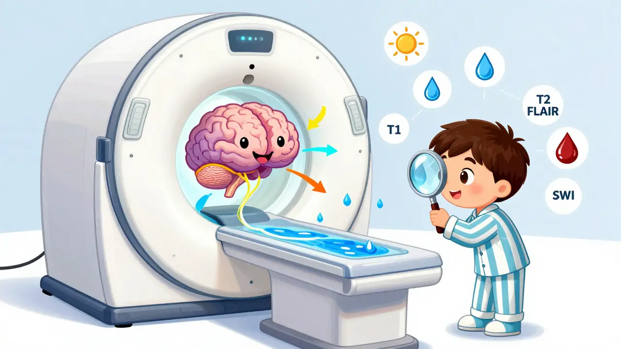
Unlike X-rays or CT scans, MRI doesn’t use radiation. Instead, it uses powerful magnets and radio waves to map the water content in your brain tissue. Different types of tissue - gray matter, white matter, cerebrospinal fluid, blood vessels - hold water differently. The machine picks up these differences and turns them into detailed images.
Modern brain MRIs are done on machines with 1.5T or 3.0T magnetic strength. The 3.0T machines give sharper images, especially for tiny structures like cranial nerves. A typical scan takes 30 to 45 minutes. You lie still in a narrow tube while the machine makes loud knocking sounds. It’s not painful, but it can feel claustrophobic. If you’re anxious, talk to your doctor about sedation options.
The real power of MRI comes from its ability to show soft tissue contrast - about 100 times better than CT scans. That means it can spot subtle changes in the brain long before they show up on other tests. That’s why it’s the gold standard for diagnosing conditions like multiple sclerosis, brain tumors, and early strokes.
The Five Key MRI Sequences You Need to Know
Doctors don’t rely on just one image. They look at a series of different scans, called sequences. Each one highlights different things. Here are the five you’ll see most often:
- T1-weighted: This is the anatomical map. Fat and some types of tissue appear bright white. Cerebrospinal fluid (CSF) looks dark. It’s great for seeing the shape of brain structures - ventricles, brainstem, cortex. If your brain looks shrunken on T1, that’s atrophy, which can happen with aging or dementia.
- T2-weighted: Water shows up bright here. Swelling, inflammation, and most lesions glow white. But here’s the catch: CSF also looks bright. That’s why you can’t tell if a bright spot is a lesion or just fluid without looking at another sequence.
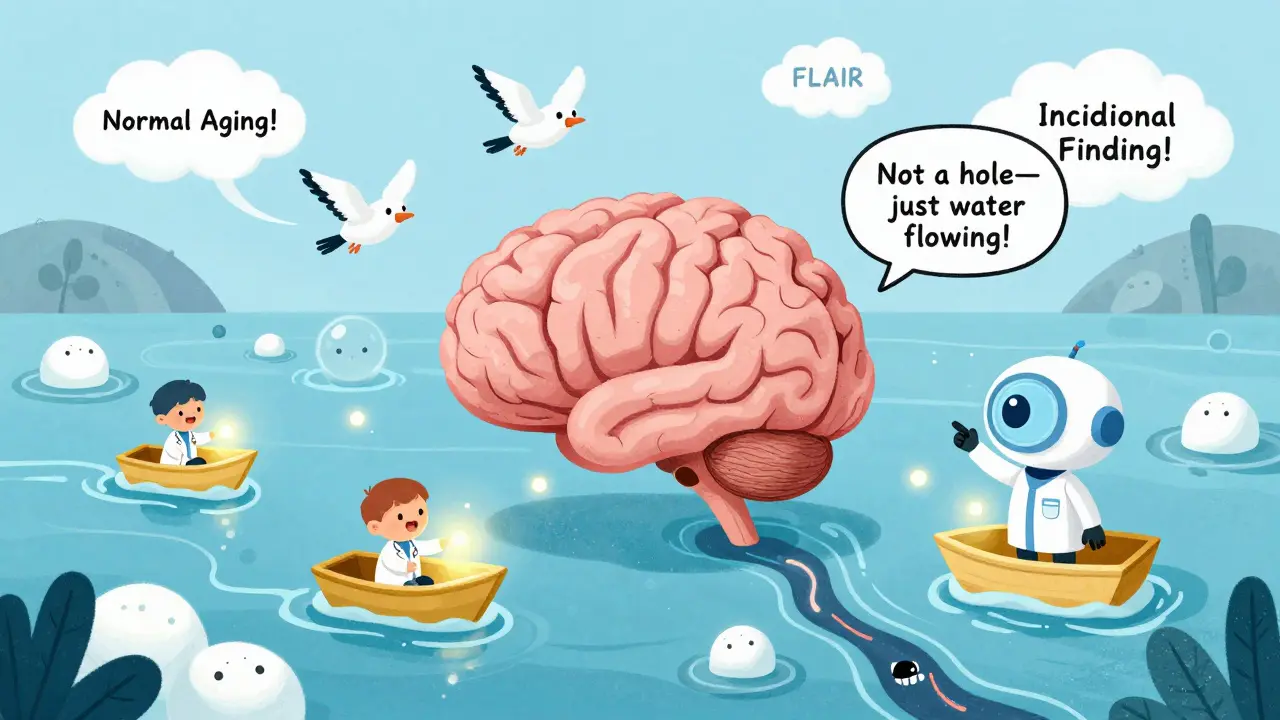
- FLAIR: This is T2 with the CSF signal turned off. It’s the most important sequence for spotting brain disease. MS plaques, small strokes, and infections show up as bright spots against a dark background. If you see bright dots around the ventricles on FLAIR, they’re likely tiny blood vessel changes - common in older adults and not always serious.
- Diffusion-weighted imaging (DWI): This picks up acute strokes within minutes. When brain cells die from lack of oxygen, water can’t move freely. DWI shows that restriction as bright spots. If DWI is normal, an acute stroke is very unlikely. ADC maps confirm it - if the numbers are below 600 x 10^-6 mm²/s, it’s an active stroke.
- SWI or Gradient Echo: This finds blood. Tiny bleeds, even those too small to see on CT, show up as dark spots. These are common in older people with high blood pressure or after head injuries. Finding them doesn’t always mean something’s wrong - but they’re clues.
Doctors always compare these sequences together. A spot that’s bright on T2 and FLAIR but dark on DWI? Probably old. Bright on DWI and FLAIR? That’s a new stroke. Bright on SWI? Could be a bleed. The pattern tells the story.
Common Findings - And What They Really Mean
Not every abnormality on an MRI is dangerous. Here are five findings you’re likely to hear about:
- White matter hyperintensities: These are bright spots on FLAIR or T2, often near the ventricles. They’re called “leukoaraiosis.” In people over 60, they’re extremely common - up to 90% of those over 70 have them. They’re linked to small vessel disease, high blood pressure, or aging. Unless they’re massive or worsening fast, they’re usually not a cause for alarm.
- Small lacunar infarcts: These are tiny (3-5mm) strokes in deep brain areas like the thalamus or basal ganglia. They often cause no symptoms. Many people live with them without knowing. But if you have several, it raises your risk for future stroke or dementia.
- Brain atrophy: The brain naturally shrinks with age. But if it’s happening faster than expected - especially in the hippocampus - it could point to Alzheimer’s or other dementias. The key is comparing it to your age group. A 75-year-old with mild atrophy is normal. A 50-year-old with severe atrophy isn’t.
- Incidental meningiomas or schwannomas: These are slow-growing, usually benign tumors. A 2mm vestibular schwannoma (on the hearing nerve) might be found on an MRI done for dizziness. Most never grow or cause problems. Doctors often just watch them with repeat scans.
- Flow voids: These are dark lines on T2 or FLAIR. They’re not lesions - they’re blood vessels. Beginners often mistake them for damage. Experienced radiologists know they’re normal. Look for symmetry. If both sides have the same dark lines, it’s just blood flow.
One of the biggest mistakes is overinterpreting. A 2021 JAMA Neurology study found that only 1.3% of brain MRIs done for routine headaches without neurological symptoms showed anything serious. Most people get scared by normal aging changes.
When MRI Is the Best Choice - And When It’s Not
MRIs aren’t always the right first test. In trauma, stroke, or emergency situations, CT is faster - done in 5 minutes versus 45 for MRI. If someone’s having a stroke and needs clot-busting drugs, waiting for an MRI could cost them brain function.
Here’s when MRI wins:
- Multiple sclerosis - MRI finds plaques with 97% accuracy, while CT misses 35%.
- Early ischemic stroke - DWI detects changes in under 30 minutes. CT often looks normal for hours.
- Brain tumors - MRI shows size, location, and how it’s affecting surrounding tissue better than any other test.
- Seizure disorders - Especially temporal lobe epilepsy, where MRI can find subtle scarring invisible on CT.
- Chronic conditions like dementia or Parkinson’s - MRI helps rule out other causes and track progression.
CT still wins for:
- Acute head trauma - It’s fast and shows skull fractures and bleeding clearly.
- Patients with metal implants - Pacemakers, cochlear implants, or certain aneurysm clips make MRI unsafe.
- Emergency settings - If you’re unstable, you get CT first.
What Your Doctor Isn’t Telling You
There’s a lot of pressure to order MRIs - from patients, from fear, from defensive medicine. But not every headache needs a scan. The American College of Radiology says MRI is “usually not appropriate” for uncomplicated migraines.
Doctors are trained to look for “red flags”: sudden severe headache (thunderclap), new seizures, weakness on one side, vision loss, confusion, or fever. If you have those, MRI is urgent. If you have a 10-year history of migraines with the same pattern? Probably not.
Also, MRI can’t tell you how old a lesion is without contrast or advanced techniques. A bright spot on FLAIR could be from a stroke that happened yesterday - or 10 years ago. That’s why your doctor will ask about your symptoms, not just look at the scan.
And yes, MRIs are expensive - $1,200 to $3,500 in the U.S. In rural areas, only 42% of hospitals have one. That’s why access isn’t equal. But in places where it’s available, it’s often the only way to see what’s really going on inside the brain.

How Radiologists Read an MRI - A Simple System
Experts don’t just glance. They follow a routine. Here’s how they do it:
- Start with the midline. Is the brain centered? Is the third ventricle symmetrical? Shifts can mean swelling or a mass.
- Check the ventricles. Are they enlarged? That’s atrophy. Are they compressed? That’s a tumor or bleeding.
- Look at the basal ganglia and thalamus. Bright spots here? Old small strokes.
- Scan the cortex. Is there swelling? Is the gray-white border sharp? Blurry borders can mean infection or inflammation.
- Check the cerebellopontine angle. This tiny space behind the ear is where acoustic neuromas hide. Miss it, and you miss a 2mm tumor.
- Review FLAIR and DWI together. That’s where most pathology hides.
One radiologist I spoke with said: “If you check the cerebellopontine angle on every scan, you’ll find something unusual once every 50 scans. Most of the time, it’s nothing. But sometimes, it’s everything.”
What’s Next for Brain MRI?
Technology is moving fast. 7.0T MRI machines - still mostly in research - can show brain layers thinner than a human hair. AI is cutting scan times in half without losing detail. Quantitative MRI, which measures actual tissue properties like water content or blood flow, is starting to replace guesswork with numbers.
By 2027, doctors may use MRI to measure myelin loss in MS or blood flow changes in early Alzheimer’s - not just see the shape, but measure the damage. That’s the future: not just pictures, but biomarkers.
For now, though, the basics still matter most. Knowing what T2, FLAIR, and DWI show can turn confusion into clarity. You don’t need to be a radiologist to understand that a bright spot isn’t always bad - and a dark spot isn’t always normal. Ask your doctor: “Which sequence shows this finding? What does it mean in context of my symptoms?” That’s how you take back control.
Is a brain MRI dangerous?
No, brain MRI is not dangerous. It doesn’t use radiation. The magnetic field is strong, but it’s not harmful. The only risks come from metal objects - pacemakers, certain implants, or metal fragments in the eye. If you have any implants, tell your doctor before the scan. Some people feel claustrophobic or anxious, but sedation is available. The noise is loud, but earplugs are always provided.
Can an MRI detect Alzheimer’s disease?
MRI alone can’t diagnose Alzheimer’s, but it can rule out other causes like tumors, strokes, or hydrocephalus. It can show brain shrinkage, especially in the hippocampus - a region tied to memory. In advanced cases, the pattern of atrophy helps support the diagnosis. Newer techniques, like measuring amyloid buildup with special MRI sequences, are being tested but aren’t standard yet.
Why do some people get abnormal MRIs but have no symptoms?
It’s more common than you think. Tiny old strokes, white matter changes, and small benign tumors often cause no symptoms. The brain has a lot of reserve. A 3mm lesion in the thalamus might be silent for decades. Doctors call these “incidental findings.” The key is whether they match your symptoms. If you feel fine and the MRI shows minor changes, it’s likely just aging - not disease.
How long does it take to get brain MRI results?
The scan itself takes 30-45 minutes. The radiologist usually interprets it within 24 to 48 hours. Your doctor will get the report and schedule a follow-up to explain it. Don’t expect results immediately after the scan - the images need expert review. Rushing the report can lead to errors.
Can I have an MRI if I have dental fillings or crowns?
Yes. Modern dental fillings, crowns, and bridges are made of non-magnetic materials and are safe for MRI. You might feel slight warmth in your mouth, but it’s harmless. Older metal fillings from the 1970s or earlier could be an issue, but those are rare. Always tell the technologist about any dental work - they’ll check the materials.
Do I need contrast dye for a brain MRI?
Not always. Many brain MRIs are done without contrast - especially for MS, stroke, or dementia. Contrast is used when doctors suspect tumors, infections, or inflammation that might be leaking blood-brain barrier. It highlights active areas. If your doctor orders contrast, it’s because they’re looking for something specific. Side effects are rare, but if you have kidney problems, let them know.
What’s the difference between a brain MRI and a spinal MRI?
A brain MRI focuses only on the brain - the cortex, cerebellum, brainstem, and ventricles. A spinal MRI looks at the spinal cord and nerve roots. They’re separate scans. You might need both if you have symptoms like numbness in your arms or legs, weakness, or bladder issues. A brain MRI won’t show spinal stenosis or disc herniations, and vice versa.
Can children have brain MRIs?
Yes. MRI is preferred for children because it doesn’t use radiation. Babies and young kids often need sedation to stay still. Many hospitals have pediatric MRI specialists who use child-friendly techniques - like playing videos or letting a parent stay in the room. It’s safe and commonly used for seizures, developmental delays, and head injuries in kids.
What to Do After Your MRI
Don’t panic over a report. Don’t Google every bright spot. Ask your doctor:
- Which sequence shows the finding?
- Is it new or old?
- Does it match my symptoms?
- Do I need another scan in the future?
Most findings are normal variants or slow-progressing changes. Only a small fraction require treatment. The goal of MRI isn’t to find something wrong - it’s to find out what’s really going on. And sometimes, the answer is: nothing serious at all.
15 Comments
Whoa, this is like a masterclass in neuro-imaging demystified. T1, T2, FLAIR, DWI - I used to think they were just alphabet soup until I read this. The part about FLAIR turning off CSF like a filter? Genius. I’ve seen those bright white dots near ventricles in my dad’s scan and panicked. Turns out they’re just the brain’s version of freckles. Mind blown. 🤯
I don’t know why people are so shocked that MRIs show ‘abnormalities’ in asymptomatic people - it’s because we’ve been sold this lie that our bodies are supposed to be perfect machines. The brain isn’t a smartphone you can just update - it’s a 3-pound organ that’s been evolving for 2 million years, and it’s got scars, glitches, and backup systems you didn’t even know existed. I had a 2mm meningioma found incidentally at 42. My neurologist said, ‘We’ll watch it.’ I said, ‘Watch it like a hawk or like a sleepy cat?’ He laughed. That’s the moment I stopped Googling. You don’t need to fix everything you find. Sometimes, you just need to live with it. And honestly? That’s the most radical thing a doctor can say anymore.
Wow so brain scans are just fancy fortune cookies now? Bright spot = bad? Dark spot = worse? LOL. I bet the radiologists are just guessing based on what they had for breakfast. Also, who the hell decided FLAIR was the most important sequence? Sounds like a brand of laundry detergent. And why do we act like DWI is some magical stroke detector? What if it’s just a glitch? Oh wait - we don’t question tech. We worship it. 🙄
White matter spots = old people. Stop panicking.
This article contains numerous factual inaccuracies and oversimplifications. For instance, the claim that MRI does not use radiation is technically correct, yet the omission of the potential biological effects of strong static magnetic fields on ion channels and neural activity is scientifically irresponsible. Furthermore, the assertion that 90% of individuals over 70 exhibit white matter hyperintensities is misleading without contextualizing comorbidities such as hypertension and diabetes, which are not merely age-related but lifestyle-driven pathologies. This kind of content promotes medical illiteracy under the guise of accessibility.
THIS. IS. EVERYTHING. 🥹 I showed this to my mom after her scan and she finally stopped crying. I’ve been Googling ‘bright spot brain MRI’ for 3 days straight and almost called an ambulance. This broke it down like a Netflix doc but with way less drama. Thank you for writing this like a human who actually cares. I’m printing this out and laminating it. 💖
Another feel-good, watered-down piece of medical propaganda. You're telling people not to panic over incidental findings? That’s not reassurance - that’s institutional negligence. The real issue isn’t the scan, it’s the system that turns every minor anomaly into a crisis. If you're not going to treat it, why are you even ordering it? You’re just creating anxiety for billing purposes. And don’t get me started on ‘AI cutting scan times in half’ - that’s code for ‘we’re replacing radiologists with algorithms that don’t understand context.’ Wake up. This isn’t medicine. It’s surveillance capitalism with a stethoscope.
So let me get this straight - we’re supposed to just ignore brain lesions because they’re ‘common’? What’s next? ‘Oh, that tumor? Probably just stress.’ I’ve seen people die from ‘incidental’ findings that were ignored. This article is dangerous. You’re giving people permission to be lazy about their health. If you see something weird on an MRI, you don’t shrug. You investigate. Period. This isn’t ‘normal aging’ - it’s negligence dressed up as wisdom.
Everyone’s so obsessed with the scans they forget the person. My sister had a 3mm lesion. Doctors kept saying ‘it’s probably nothing.’ She had a seizure six months later. The lesion was a low-grade glioma. You can’t just say ‘it’s common’ and move on. If you’re going to show people images, you owe them the truth - not a pep talk. This article is a disservice to anyone who’s ever been told ‘don’t worry’ when they should’ve been told ‘let’s run more tests.’
thx for this!! i had an mri last month for dizziness and the report said 'mild white matter changes' and i thought i was dying. now i feel way better. i think i just needed someone to say 'hey, your brain is just old like my coffee maker' lol. also the part about flow voids? mind blown. i thought they were holes in my brain 😅
It’s fascinating how we treat the brain as a machine to be fixed, rather than a living system that adapts. A bright spot isn’t a flaw - it’s a story. Maybe it’s a micro-stroke from a night of stress. Maybe it’s the echo of a childhood fall. Maybe it’s just the brain’s way of saying, ‘I’ve been here a long time, and I’m still working.’ We don’t need more scans. We need more curiosity. Less fear. More patience.
This is a well-structured educational resource that aligns with current clinical guidelines. The emphasis on sequence interpretation and differential diagnosis is commendable. However, the casual tone undermines the gravity of neurological pathology. Medical communication must balance clarity with responsibility. I recommend incorporating standardized terminology from the ACR Appropriateness Criteria to ensure consistency across patient populations.
Man, I wish I had this 5 years ago. My wife had that ‘bright spot’ thing and we were up all night reading scary blogs. This is the kind of stuff you wish your doctor just handed you instead of saying ‘we’ll talk next week.’ I’m sharing this with everyone I know. Also, the cerebellopontine angle tip? Gold. I’m gonna start checking that on every scan I see 😎
As someone who grew up in a village with no MRI machine, I’ve seen what happens when you don’t have access. People wait until they’re dying before they get scanned. This article is a gift - but only if it reaches the right people. Let’s not make this knowledge a luxury. We need community health workers teaching this stuff in local languages. Knowledge shouldn’t be gated by WiFi or hospital budgets. 🙏
How quaint. You’ve reduced the complexity of neuroimaging to a BuzzFeed listicle. The five sequences? Please. Real radiologists spend years mastering the interplay of relaxation times, field inhomogeneities, and artifact suppression. You’re not empowering patients - you’re feeding them soundbites that make them think they can interpret scans better than their own neurologist. This isn’t education. It’s epistemic arrogance disguised as accessibility.